
Strongly pleochroic: Yellow to lavender to red
Cobalt phosphate
Anhydrous cobalt phosphate. Co3(PO4)2
The refractive index is rather variable n = c. 1.65 to c. 1.81
Under the microscope, the pigment tends to be in the form of irregularly-shaped, plate-like particles and broken crystal forms, which may include gas bubbles. The particles range in size from 4 to 50 micrometres. It is very clearly pleochroic and shows a variety of colours from red to yellow to pale lavender. Between crossed polars the extinction is dispersed and consiquently the interference colours are anomalous; it appears to show second order green and possibly third order colours. Many particles do not extinguish but merely change colour as the stage is rotated. In non-plane-polarised light, the particles vary in colour from bluish-red to violet and become strongly red with the Chelsea filter.
The method of preparing this pigment was first published in 1859. It is made by precipitating a soluble cobalt salt with disodium phosphate, washing the precipitate and then heating it strongly.
This pigment, often called cobalt violet dark, is extremely stable; it is unaffected by most reagents and sunlight. It is reddish violet in colour, is transparent in oil, has low tinting power, and exhibits pronounced purple/violet metameric effects. It can be used in all techniques. It was not listed by Winsor and Newton in 1896, but was listed in 1928.
Strongly pleochroic: Yellow or white to dark blue
Vivianite
Refractive index 1.579 to 1.636
Fe3P2O8 . 8H2O
Under the polarising microscope a sample supplied by Dr. Kremer was seen to be composed of particles up to 60 micrometres long. Vivianite shows good cleavage and most of the particles were flat plates sometimes showing parallel cracks. In meltmount these flakes were nearly colourless with markedly yellow edges. Most of the particles had Becke lines that moved out but in a few instances they moved inwards (this is probably related to the form of the edges of the particles). Extinction was definite and very sharp for the cleavage flakes. The birefringence was low – few particles exhibited anything but a first order grey or white. Some plates showed slight pleochroism changing from white or yellowish to faint blue. Another, less common type of particle tended to be noticeably longer than it was wide, and had striations running down its length. Such particles showed marked pleochroism from a deep blue to white or yellow. Extinction of the striated particles was mostly undulose or oblique with anomalous colours: pinks and bronze colours appearing at the position where extinction was expected, but some particles extinguished straight. The blue particles did not show pink or red with the Chelsea filter.
Vivianite is formed by the action of phosphorous-containing solutions on iron compounds and is found in, among other places, the forests in Bavaria. according to Winchell, the mineral is colourless when fresh, but changes to blue and finally brown on oxidising. The change to blue is instantaneous when the mineral is ground, as a result of oxidation. (Winchell, A. N. Elements of Optical Mineralogy Pt II, 3rd Ed. 1933, p. 126.) The colour of the dry pigment is a dull slaty blue. It has been found in organic phosphate-rich environments, often associated with bones, decaying wood and other organic remains.
The pigment is not very strongly coloured and, depending on the medium and the varnish, can appear a greyish black. It is mainly thought of as occurring on German objects and wall paintings that can be dated between 1150 and 1235, but it has also been found on 12th and 13th century paintings at Winchester and has been tentatively identified from Anglo Saxon stone sculpture at York. The authors have been told that it was found on 18th century Bavarian window shutters. Originally, it may have come into use because there were few blue pigments available to artists at the time. Egyptian blue was probably no longer being made, and ultramarine was only just beginning to be used. Nevertheless, vivianite does not appear to have been regarded merely as a cheap substitute for ultramarine, because the two pigments were found side by side on the lectern that is preserved at the City Church of Freuenstädter and which dates from c. 1150. (Richter, Ernst-Ludwig, Seltene Pigmente im Mittelalter, Zeitchrift für Kunsttechnologie und Konservierung, Vol I No. 2, 1988.)
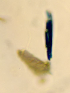

the stage has been rotated 90°compared with the previous image.
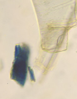
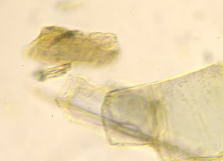
the stage has been rotated 90° compared with image above
Note the parallel cracks in the large cleavage fragment on the right
and that it has hardly changed in colour
Mactaggart, P. & Mactaggart, A. (June 2007) ‘Strongly Pleochroic’ In: Pigment ID using Polarised Light Microscopy from: https://academicprojects.co.uk/strongly-pleochroic/
